Home » Uncategories » Anatomy Of Chest / Surgical Anatomy Of The Chest Wall Thoracic Key - The thorax has two major openings:
Friday, 25 June 2021
Anatomy Of Chest / Surgical Anatomy Of The Chest Wall Thoracic Key - The thorax has two major openings:
Anatomy Of Chest / Surgical Anatomy Of The Chest Wall Thoracic Key - The thorax has two major openings:. Chest pain has many possible causes, all of which need medical attention. Computed tomography (ct) of the chest can detect pathology that may not show up on a conventional chest radiograph(1). The circulatory system does most of its work. The dominant muscle in the upper chest is the pectoralis major. The chest anatomy includes the pectoralis major, pectoralis minor and the serratus anterior.
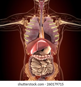
Three dimensional view of the female reproductive system, full frontal view. The right side of the heart is deflected anteriorly, and the left side is deflected posteriorly. The superior thoracic aperture found superiorly and the inferior thoracic aperture. Anatomically, the heart is located in the anterior thoracic cavity; Chest a man's chest — like the rest of his body — is covered with skin that has two layers.
Flail Chest Background History Of The Procedure Problem from img.medscapestatic.com The muscles of the chest develop from the somites found in the mesoderm. Chest a man's chest — like the rest of his body — is covered with skin that has two layers. The pectoralis major and the pectoralis minor, known collectively as your pecs. This thoracic and pulmonary anatomy tool is especially designed for students of anatomy (medical and paramedical studies). The chest is made up primarily of two muscles: (1) the pectoralis major, and (2) the pectoralis minor. And flexibility to aid in the functional process of respiration. This chapter is an abbreviated review of thoracic anatomy as seen on chest radiographs and computed tomography (ct) of the chest.
About the 6th week, the somites differentiate into the sclerotomes and the dermatomyotomes.
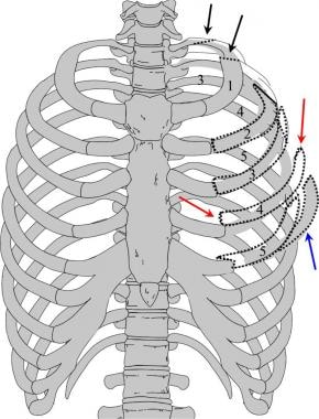
Download my two educational text books for free using this link: You will also find the xiphoid process, 10th rib, the apex of the heart, the coronary vein of the heart. And flexibility to aid in the functional process of respiration. The chest wall is a complex system that provides rigid protection to the vital organs such as the heart, lungs, and liver; The chest is the area of origin for many of the body's systems as it houses organs such as the heart, esophagus, trachea, lungs, and thoracic diaphragm. This chapter is an abbreviated review of thoracic anatomy as seen on chest radiographs and computed tomography (ct) of the chest. Anatomy of the thorax, heart, abdomen and pelvis recommended text gray's anatomy for students. Sternocleidomastoid muscle clavicle and ribs anatomy muscle anatomy chest sternocleidomastoid ribs anatomy chest muscles anatomy thorax rib muscles chest muscles chest anatomy illustration. The chest anatomy includes the pectoralis major, pectoralis minor and the serratus anterior. Three dimensional view of the female reproductive system, full frontal view. It is enclosed by the ribs, the vertebral column, and the sternum, or breastbone, and is separated from the abdominal cavity (the body's largest hollow space) by a muscular and membranous partition, the diaphragm. (1) the pectoralis major, and (2) the pectoralis minor. Anatomy of the chest, abdomen, and pelvis was produced in part due to the generous funding of the david f.
Three dimensional view of the female reproductive system, full frontal view. Thoracic cavity, also called chest cavity, the second largest hollow space of the body. Learn about each of these muscles, their locations, functional anatomy and exercises for them. Stability to arm and shoulder movement; About the 6th week, the somites differentiate into the sclerotomes and the dermatomyotomes.
Nerves Of The Chest And Upper Back from www.innerbody.com Chest bone, ribs, lung, heart, xiphoid process, sternum anatomy. Radiology basics of chest ct anatomy with annotated coronal images and scrollable axial images to help medical students and junior doctors learning anatomy. A good radiologist knows the anatomy because knowing where structures normally live and recognizing the location of an abnormality helps to make or narrow the differential diagnosis. #anatomy of the chest and stomach. Learn about each of these muscles, their locations, functional anatomy and exercises for them. The superior thoracic aperture found superiorly and the inferior thoracic aperture. Anatomy of the thorax, heart, abdomen and pelvis recommended text gray's anatomy for students. The chest wall is a complex system that provides rigid protection to the vital organs such as the heart, lungs, and liver;
12 photos of the anatomy of the chest and stomach.
(1) the pectoralis major, and (2) the pectoralis minor. The chest is the area of origin for many of the body's systems as it houses organs such as the heart, esophagus, trachea, lungs, and thoracic diaphragm. The first step in understanding thorax anatomy is to find out its boundaries. Computed tomography (ct) of the chest can detect pathology that may not show up on a conventional chest radiograph(1). Chest pain has many possible causes, all of which need medical attention. Muscles of the chest and their functions you have two mighty muscles on both sides of your chest: A good radiologist knows the anatomy because knowing where structures normally live and recognizing the location of an abnormality helps to make or narrow the differential diagnosis. Thoracic cavity, also called chest cavity, the second largest hollow space of the body. The circulatory system does most of its work. In insects, crustaceans, and the extinct trilobites, the thorax is one of the three main divisions of the creature's body, each of which is in turn composed of multiple segments. Download my two educational text books for free using this link: Swensen fund for innovation in teaching. #anatomy of the chest and stomach.
Chest a man's chest — like the rest of his body — is covered with skin that has two layers. The first step in understanding thorax anatomy is to find out its boundaries. This page provides an overview of the chest muscle group. Anatomically, the heart is located in the anterior thoracic cavity; A good radiologist knows the anatomy because knowing where structures normally live and recognizing the location of an abnormality helps to make or narrow the differential diagnosis.
Human Chest Anatomy Images Stock Photos Vectors Shutterstock from image.shutterstock.com Browse 6,409 chest anatomy stock photos and images available, or search for human anatomy to find more great stock photos and pictures. Download my two educational text books for free using this link: The thorax has two major openings: It is important to remember the position and orientation of the heart when placing a stethoscope on the chest of a patient and listening for heart sounds, and also when looking at images taken from a midsagittal perspective. Three dimensional view of the female reproductive system, full frontal view. The muscles of the chest develop from the somites found in the mesoderm. The superior thoracic aperture found superiorly and the inferior thoracic aperture. Here, we break down the anatomy of your chest muscles.
(1) the pectoralis major, and (2) the pectoralis minor.
The chest is the area of origin for many of the body's systems as it houses organs such as the heart, esophagus, trachea, lungs, and thoracic diaphragm. Stability to arm and shoulder movement; Anatomy of the thorax, heart, abdomen and pelvis recommended text gray's anatomy for students. Three dimensional view of the female reproductive system, full frontal view. Diseases of the chest and chest abnormalities make up a significant portion of a physician's daily practice. Anatomy of the chest and the lungs: The thorax has two major openings: The pectoralis major and the pectoralis minor, known collectively as your pecs. Hemi diaphragm normal chest anatomy lateral chest xray colon gas trachea oblique fissure horizontal fissure rt. This work was supported in part by the kaplow family fund, yale school of medicine. The right side of the heart is deflected anteriorly, and the left side is deflected posteriorly. You will also find the xiphoid process, 10th rib, the apex of the heart, the coronary vein of the heart. The chest anatomy includes the pectoralis major, pectoralis minor and the serratus anterior.
0 Response to "Anatomy Of Chest / Surgical Anatomy Of The Chest Wall Thoracic Key - The thorax has two major openings:"


0 Response to "Anatomy Of Chest / Surgical Anatomy Of The Chest Wall Thoracic Key - The thorax has two major openings:"
Post a Comment